Image guided surgery of the Brain and Spine
The trend in neurosurgery is characterized by minimizing of surgical trauma and the improvement in visibility of the surgical field. The use of neuroendoscopy during neurosurgical procedures is the modern triumph.
The endoscope is used during trans-nasal transphenodial removal of pituitary adenoma, during the insertion of drainage valves for hydrocephaly, to perform third ventriculostomy to remove skull base tumors in combination with microneurosurgery.
Another method for the precise location of the position of a tumor is stereotactic. Special equipment (special stereotactic frame and software) and with the guidance of CT or MRI offers the possibility of determining the damage to the millimeter.
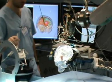
Newer techniques however use special computer assisted equipment and robotic arms or special microscopes to access and remove respective tumors.
The abovementioned techniques can be applied to non-surgically accessible tumors which cannot be accessed and resected (eg tumors of brainstem and hypothalamus) to take a biopsy or brachytherapy with placement of microcatheter for radioisotope infusion, radiolabelled antibodies chemotherapeutic drugs or genetically engineered substances.
Over the past three decades, advances in surgical neuro oncology have been made in two sectors: the equipment which allows minimally invasive surgery and the quality and variety of imaging methods. The two sectors are now combined defining the beginning of image-assisted minimally invasive surgery.

Neuronavigation
Virtual imagery, or otherwise neuro navigation can be used before surgery to plan or even to carry out a test surgery. It can also be used for planning surgical approach. It can also be used in real time to record the progression of the surgery and to see beyond the visual field to protect the patient from the destruction of structures beneath the surface invisible to the surgical microscope.
There are two basic methods of using images to navigate in three dimensions. For micro millimeter accuracy an external stereotactic ring is used, which is fixed to the skull and used as a reference, this developed into a system without a ring, which allows recording on the patient's skin or bone and can perceive changes in real time. Therefore, a series of cameras continuously record the positions of the patient and surgical instruments and play them on screen in three dimensions in relation to the tumor of patient, the blood vessels and the healthy structures of the brain with a delayed measurement of only a few milliseconds.
Intraoperative imaging
The construction of a virtual brain (and tumor) is a big step forward, but the brain is not a static organ. It begins to move as soon as the skull is opened and continues to move as cerebrospinal fluid (CSF) continues to decrease during surgery.
Future applications
The sensitivity of magnetic resonance imaging (MRI) to changes in temperature is an important element in typesetting of new therapeutic methods for brain tumors. This capability allows the neurosurgeon to record thermal variations in microsurgical techniques, such as laser treatment, radiofrequency therapy, focused ultrasound techniques and cryosurgery. The focused ultrasound is a very attractive option because it does not need the surgeon to perform craniotomy, or to dissect the skin to thermally remove a tumor.
The trend in neurosurgery is characterized by minimizing of surgical trauma and the improvement in visibility of the surgical field. The use of neuroendoscopy during neurosurgical procedures is the modern triumph.

Microsurgery with neuro Navigation
The endoscope is used during trans-nasal transphenodial removal of pituitary adenoma, during the insertion of drainage valves for hydrocephaly, to perform third ventriculostomy to remove skull base tumors in combination with microneurosurgery.
Another method for the precise location of the position of a tumor is stereotactic. Special equipment (special stereotactic frame and software) and with the guidance of CT or MRI offers the possibility of determining the damage to the millimeter.
Newer techniques however use special computer assisted equipment and robotic arms or special microscopes to access and remove respective tumors.
The abovementioned techniques can be applied to non-surgically accessible tumors which cannot be accessed and resected (eg tumors of brainstem and hypothalamus) to take a biopsy or brachytherapy with placement of microcatheter for radioisotope infusion, radiolabelled antibodies chemotherapeutic drugs or genetically engineered substances.
Over the past three decades, advances in surgical neuro oncology have been made in two sectors: the equipment which allows minimally invasive surgery and the quality and variety of imaging methods. The two sectors are now combined defining the beginning of image-assisted minimally invasive surgery.
Neuronavigation
Virtual imagery, or otherwise neuro navigation can be used before surgery to plan or even to carry out a test surgery. It can also be used for planning surgical approach. It can also be used in real time to record the progression of the surgery and to see beyond the visual field to protect the patient from the destruction of structures beneath the surface invisible to the surgical microscope.
There are two basic methods of using images to navigate in three dimensions. For micro millimeter accuracy an external stereotactic ring is used, which is fixed to the skull and used as a reference, this developed into a system without a ring, which allows recording on the patient's skin or bone and can perceive changes in real time. Therefore, a series of cameras continuously record the positions of the patient and surgical instruments and play them on screen in three dimensions in relation to the tumor of patient, the blood vessels and the healthy structures of the brain with a delayed measurement of only a few milliseconds.
Intraoperative imaging
The construction of a virtual brain (and tumor) is a big step forward, but the brain is not a static organ. It begins to move as soon as the skull is opened and continues to move as cerebrospinal fluid (CSF) continues to decrease during surgery.
Future applications
The sensitivity of magnetic resonance imaging (MRI) to changes in temperature is an important element in typesetting of new therapeutic methods for brain tumors. This capability allows the neurosurgeon to record thermal variations in microsurgical techniques, such as laser treatment, radiofrequency therapy, focused ultrasound techniques and cryosurgery. The focused ultrasound is a very attractive option because it does not need the surgeon to perform craniotomy, or to dissect the skin to thermally remove a tumor.
No comments:
Post a Comment